Your Subtitle text

Urinary Incontinence

Urinary incontinence is more common in women than in men.  Some women have the false belief that urinary incontinence is a normal part of aging and that nothing can be done to correct it.  Urinary incontinence can often be treated.  It is important to discuss any urinary leakage with your doctor.  Proper diagnosis and treatment may correct your problems and reduce your symptoms.

There are different types of incontinence and there are many possible causes.  These include infection, damage to organs, muscular disorders and medications.  A number of steps may be needed to determine the cause.  There are many options for treatment, depending on the cause of your problem.   Treatments can include behavioral changes such as bladder retraining and pelvic muscle exercises (Kegels), medication, special devices, and surgical options.

Urinary incontinence is a common problem.  If you have symptoms, or if they affect your activities of daily living, tell your doctor.  In most cases this can be treated with success.

Pelvic Floor Exercises (Kegels)

These exercises are performed to improve the tone of the perineal muscles and pelvic organ support, promote postpartum healing and comfort, prevent urinary incontinence or regain bladder control.  They may be done in any position.

Tightly contract the muscles surrounding the vagina with an internal “pulling up”.  Release the muscles and notice the relaxation.  Again contract tightly, hold and released.  Do not tighten the stomach muscles during this exercise.  Breathe normally throughout.  If it is difficult to get the feeling of this exercise, try stopping and starting the flow if urine while urinating.

When first starting the exercise, perform five in a series, holding each muscle contraction for five seconds, then release.  Perform 50 a day for the rest of your life.


Osteoporosis

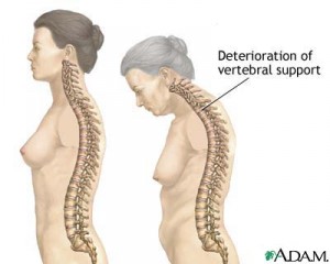
Osteoporosis is a disease that causes bones to become thin and break easily. Since the consequences of a fracture are so great, it is important to know that osteoporosis can be prevented by proper nutrition, exercise, healthy lifestyle choices, early diagnosis and medication when needed.

Important risk factors include: female, Caucasian, advanced age, history of bone fracture, a small thin frame, family history of osteoporosis, early menopause, removal of the ovaries, a low calcium diet, lack of exercise, eating disorders, certain medicines (steroids and anticonvulsants), alcohol and tobacco use.

Bone healthy actions need to begin in childhood and continue throughout life.  Before menopause, it is important for women to have regular menstrual cycles. The hormone estrogen regulates the menstrual cycle and promotes bone health.  In women, excessive thinness often disrupts normal menstrual cycles.  Eating disorder can lead to osteoporosis.  If your cycles are infrequent or irregular, or if you think you may have an eating disorder, it is imperative to speak with your doctor.

While it is natural to lose some bone as you age, it is not natural to develop osteoporosis, experience painful fractures or to lose more than 1-1/2 inches of height.

A medical test that measures the amount of bone is the best way to detect osteoporosis.  A bone densitometer is one such test that uses small amounts of x-ray to measure the amount of bone mineral.  The amount of bone mineral relates directly to bone strength.  The test is painless and usually takes less than ten minutes.  A result called a T-score will be sent to your doctor.


Mammogram

A mammogram is an X-ray test of the breasts (mammary glands) used to screen for breast problems, such as a lump, and whether a lump is fluid-filled (a cyst) or a solid mass.

A mammogram is done to help screen for or detect breast cancer. Many small tumors can be seen on a mammogram before they can be felt by a woman or her health professional. Cancer is most easily treated and cured when it is discovered in an early stage. Mammograms do not prevent breast cancer or reduce a woman’s risk of developing cancer. But regular mammograms can reduce a woman’s risk of dying from breast cancer by detecting a cancer when it is more easily treated.

Experts differ in their recommendations about when or how often women should have mammograms.

How It Is Done

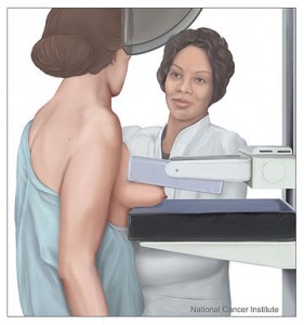
A mammogram is done by a radiology technologist or mammogram technologist. The X-ray pictures (mammograms) are interpreted by a doctor who specializes in evaluating X-rays (radiologist).

You will need to remove any jewelry that might interfere with the X-ray picture. You will need to take off your clothes above the waist, and you will be given a cloth or paper gown to use during the test. If you are concerned about an area of your breast, show the technologist so that the area can be noted.

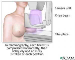
You usually stand during a mammogram; sometimes you may also be asked sit or lie down, depending upon the type of X-ray equipment used. One at a time, your breasts will be placed on a flat plate that contains the X-ray film. Another plate is then pressed firmly against your breast to help flatten out the breast tissue. Very firm compression is needed to obtain high-quality pictures. You may be asked to lift your arm or use your hand to hold your other breast out of the way. For a few seconds while the X-ray picture is being taken, you will need to hold your breath. Usually at least two pictures are taken of each breast: one from the top and one from the side.

You may be in the mammogram clinic for up to an hour; the mammogram itself takes about 10 to 15 minutes. You will be asked to wait (usually about 5 minutes) until the X-rays are developed, in case repeat pictures need to be taken. In some clinics and hospitals, X-ray pictures can be viewed immediately on a computer screen (digitally).

How It Feels

A mammogram is often uncomfortable but rarely extremely painful. If you have sensitive or fragile skin, or a skin condition, let the technician know before you have your exam. If you have menstrual periods, the procedure is more comfortable when done within 2 weeks after your period has ended.

The X-ray plate will feel cold when you place your breast on it. Having your breasts flattened and squeezed is usually uncomfortable. But it is necessary to flatten out the breast tissue to obtain the best pictures.


Abnormal Pap test Results

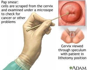
The Pap test is a way to find cell changes on the cervix.  If a Pap test shows these changes, the result will be called abnormal.  In some cases, these abnormal cells may lead to cancer. You may need treatment. In most cases, the treatment will be performed in the doctor’s office with good results.

The Pap test or Pap smear is an important part of the women’s health care.  It tells the doctor if there are any abnormal cells on the cervix.  Some abnormal cells may be precancer, or rarely, cancer.  Precancer is when there are changes in the cells that may, but do not always, become cancer if left untreated.

An abnormal Pap test is usually caused by an infection such as human papilloma virus (HPV) or types of vaginal irritation or cellular changes.  In women who smoke, however, HPV infections are more likely to cause cell changes than in women who do not smoke.  Smoking lowers the ability of the immune system to get rid of the HPV.  Quitting smoking markedly improves the immune system, making it more likely that the Pap will return to normal without treatment.

A woman who receives an abnormal Pap test result may need further testing.  Further testing methods such as colposcopy and biopsy can help identify the reason for the abnormal test result.

Treatment of cervical changes depends on the severity of the problem.

If you are concerned about abnormal Pap test results, talk to your doctor.  Most problems that cause abnormal Pap test results, when found early, can be treated.  Routine exams and Pap tests are the best was to find cervical problems.


Colposcopy

Colposcopy is a way of looking at the cervix through a special magnifying device called a colposcope.  It shines a light onto the vagina and cervix.  A colposcope can enlarge the normal view allowing the doctor to find problems that cannot be seen by the eye alone.

Colposcopy is done when a Pap test shows abnormal changes.  It provides more information about the abnormal cells.  It is done like a Pap test in a doctor’s office.  The procedure is best done when a woman is not having her menstrual cycle. For at least 24 hours before the test it is best not to douche, use tampons, use vaginal medications or have intercourse.  A mild solution will be applied to your cervix and vagina making abnormal areas easier to see.  You may feel a slight burning.

If abnormal areas are seen, a biopsy may need to be done.  A small piece of tissue will be removed with a special device. Cells also may be taken from the canal of the cervix by a scraping called endocervical curettage (ECC).  If a biopsy or scraping was done, it will be sent to a lab for further study.

If you have a colposcopy with biopsy, you may have some vaginal bleeding, and/or a dark discharge for a few days.  While the cervix heals, do not put anything in your vagina for at least one week.  You should call your doctor right away if you experience heavy bleeding using more than one sanitary pad per hour, severe lower abdominal pain, fever, chills or foul smelling discharge.

Follow up with your doctor for the results of your colposcopy.

Laparoscopy

Laparoscopy can be useful in diagnosing and treating many gynecological problems.  It has taken the place of surgery in some cases with fewer problems and shorter recovery.

A laparoscope is a small telescope that is inserted into the abdomen through a small incision.  It brings light into the abdomen so the doctor can see inside.

There are many reasons why your doctor may use laparoscopy.  For example, it can be used to diagnose and treat endometriosis, ovarian cysts, ectopic pregnancy and fibroids.

Laparoscopy is usually done on an outpatient basis so you don’t have to stay in the hospital overnight.  Other benefits included smaller incisions and a shorter recovery time.

Anesthesia is used during the procedure.  After it is administered a small cut is made below or inside the navel.  A gas is usually put into the abdomen causing it to swell so the pelvic organs can be seen more clearly.  The laparoscope is placed through this cut.   Another cut is generally made to accommodate other instruments that may be used during the procedure.

Usually the laparoscope projects images of the surgery onto a screen, making the images larger and easier to view.   After the procedure, the instruments are removed and the gas is released.  The incisions are closed, usually with stitches that dissolve.  You could be released within a few hours, but you should plan to have someone take you home.

Common problems include: Nausea, pain around the incision sites, scratchy throat (if a breathing tube was used during anesthesia), abdominal cramps, vaginal discharge (that may last a few days), swollen abdomen, shoulder pain.  It is safe to resume normal activities as soon as you feel up to it, but avoid heavy lifting for two weeks. Resume your normal diet, but increase your fluids and fiber to avoid constipation. Talk with your doctor about when to resume intercourse.

Call your doctor immediately if you have an increase in severity in any of the above mentioned symptoms after a 24 hour period, a temperature of 101 F or greater, vomiting or increased abdominal pain.

You should have a follow up appointment in the office with your doctor within 2 weeks.